SI Joint Diagnostic Challenges
Presenting complaints mimic other causes of chronic low back pain (LBP) including the lumbar spine, and hip. Referral pain patterns for the three sources overlap, which adds to the diagnostic challenge. Imaging studies are often inconclusive which makes a thorough physical exam an important part of the differential diagnosis for LBP.
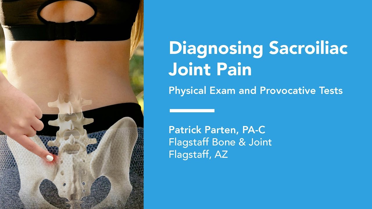
The physical exam may include palpation (PSIS, Iliac Crest, Dorsal Ligament, Sacral Sulcus), provocation tests, and the active straight leg raise test (ASLR). The Fortin Finger Test has proven to be a simple and reliable diagnostic tool to diagnose SI joint pain.2
The following tables detail the history, causes, complaints, and exacerbating or relieving activities that are commonly described by those suffering from SI joint pain.
Pain Diagram
- Pain presents in the buttock and posterior thigh
- Usually not midline
- Usually below L5
- At or lateral to PSIS
- Occasionally groin
- Secondary pain in lateral thigh, groin, and/or lateral calf

Traumatic
- Fall
- Motor Vehicle Accident
- Lifting
- Post-partum
Gradual Onset
- Laxity of the SIJ / Multiple Pregnancies
- Repetitive Forces on SIJ and Supporting Structures
- Biomechanical Abnormalities
- Leg Length Inequality
- Pelvic Obliquity/Scoliosis
- Iliac crest bone graft
- Arthritis
- Adjacent Segment Degeneration
History
- Prior Trauma
- A fall on the buttock
- Car accident (T-bone, rear-end, head-on)
- Lift/Twist
- Other
- Prior lumbar fusion
- Prior iliac bone graft harvest
- Pregnancy
Complaints
- Lower back pain
- Lower extremity pain (numbness, tingling, weakness)
- Pelvis / buttock pain
- Hip / groin pain
- Unilateral leg instability (buckling, giving away)
- Disturbed sleep patterns
- Disturbed sitting patterns (unable to sit for long periods, on one side)
- Pain when going from sitting to standing
Exacerbating Activities
Pain with Transitional Motions
- Supine to painful side
- Sit to stand
- Rolling over in bed
- Getting in & out of bed
- Pain while stationary (Sitting on affected side, prolonged standing sitting)
Unilateral Weight Bearing
- Putting on socks/shoes
- Ascending / descending stairs
- Getting in and out of car
- Prolonged walking
- 85% of Gait is Single Leg Stance
Sexual Intercourse
Relieving Activities
- Bearing weight on unaffected side.
- Lying on unaffected side
- Manual or belt stabilization
SI Joint Provocation Tests
Provocation tests have been shown to be more reliable than position/motion tests to diagnose SI joint pain.3 The following five (5) provocative tests performed in combination have a high degree of sensitivity and specificity and have been demonstrated to be generally reliable. One positive test raises suspicion, two positive tests provide fair confidence, and three positive tests provide high confidence in the diagnosis.4,5

Distraction

Thigh Thrust

Compression

Faber

Gaenslen’s
Imaging
Imaging studies for the SI joint may include plain radiographs, CT scan, MRI, or bone scan. Imaging may also identify or rule out trauma, stress fracture, inflammation, tumor, and infection.
SI Joint Injections
SI joint injections confirmed with contrast and imaging are the gold standard for the diagnosis of SI joint pain. An injection is indicated after the following:
- Negative Lumbar and Hip Exam
- Positive History
- Positive Fortin Finger Test
- Positive Physical Exam
- Positive Pain Provocation Tests
A positive injection provides >50% acute decrease in pain. If the decrease is <50% the patient may have SI joint pain, but other pain sources should be considered. Utilizing a patient pain diary to track the response is also helpful.
Treatment Options
Options for non-surgical treatment include NSAIDS, external fixation, physical therapy (4-6 weeks), intra-articular SI joint steroid injections, RF ablation. There is only low level of support in the literature for these options, but they represent a low risk to the patient.6 Patients with chronic SI joint pain lasting more than 6 months and who have failed a reasonable course of non-surgical treatment options are considered candidates for SI joint fusion.
Learn more about the SI joint fusion surgery with the Alevio’s Implants.
References
- Weksler, et al. Archives of Orthopaedic and Trauma Surgery 2007; 127, No. 10: 885-888.
- Fortin JD, Falco FJ. The Fortin finger test: an indicator of sacroiliac pain. Am J Orthop (Belle Mead NJ). 1997;26(7):477-480.
- Potter NA, Rothstein JM. Intertester reliability for selected clinical tests of the sacroiliac joint. Phys Ther 1985;65(11):1671-75.
- Laslett M, Williams M. The reliability of selected pain provocation tests for sacroiliac joint pathology. Spine 1994.
- Van der Wurff P et al. Clinical tests of the SI joint. A systematic methodological review. Part 1: Reliability. Man Ther 2000.
- Lorio M, Kube R, Araghi A. International Society for the Advancement of Spine Surgery Policy 2020 Update-Minimally Invasive Surgical Sacroiliac Joint Fusion (for Chronic Sacroiliac Joint Pain): Coverage Indications, Limitations, and Medical Necessity. Int J Spine Surg. 2020;14(6):860-895. doi:10.14444/7156.